🎯 Key Points
- Sliding filament theory: nerve impulse → Ca²⁺ release → Ca²⁺ binds troponin → tropomyosin shifts → myosin binds actin → cross-bridge cycling → sarcomere shortens; ATP needed to DETACH (not attach) myosin — this is why rigor mortis occurs after death (no ATP to release the cross-bridge)
- During contraction: I band DECREASES, H zone DECREASES, A band stays the SAME (only the overlap changes, myosin filament length doesn't)
- 206 bones in adults; Axial skeleton (80: skull, vertebral column, ribcage) + Appendicular skeleton (126: limbs, girdles)
- Joint types by mobility: Fibrous (immovable, skull sutures) → Cartilaginous (slightly movable, vertebrae) → Synovial (freely movable, limbs)
- Red/slow-twitch fibres = aerobic, fatigue slowly (endurance); White/fast-twitch fibres = anaerobic, fatigue fast (sprinting/power)
Types of Movement
- Amoeboid: pseudopodia extension (WBCs, Amoeba)
- Ciliary: cilia movement (respiratory tract, fallopian tube)
- Muscular: contraction of muscle fibres
Types of Muscle
- Skeletal (striated/voluntary): attached to bones; voluntary control; fast but tires quickly
- Smooth (unstriated/involuntary): internal organs (gut, blood vessels); slow, sustained
- Cardiac: heart only; involuntary; striated; never fatigues
Sliding Filament Theory of Muscle Contraction
- Sarcomere: functional unit (Z disc to Z disc)
- Thick filaments: myosin; thin filaments: actin, troponin, tropomyosin
- Steps: nerve impulse → Ca2+ release from SR → Ca2+ binds troponin → tropomyosin moves → myosin head binds actin → cross-bridge cycling → Z discs pulled together → sarcomere shortens
- ATP needed for cross-bridge detachment; rigor mortis = no ATP after death
- I band decreases; A band stays same; H zone decreases during contraction
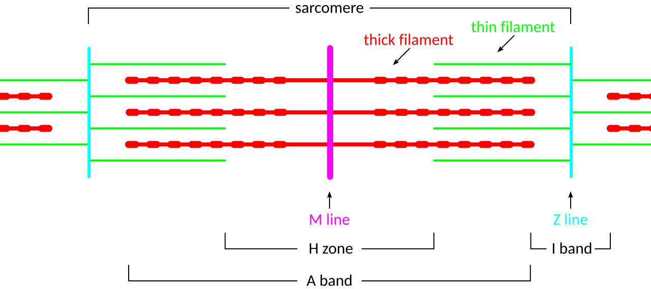
A sarcomere (Z line to Z line): during contraction the I band and H zone shorten as thin filaments slide over thick, while the A band stays the same length. Image: SlothMcCarty, CC BY-SA 3.0, via Wikimedia Commons.
Skeletal System
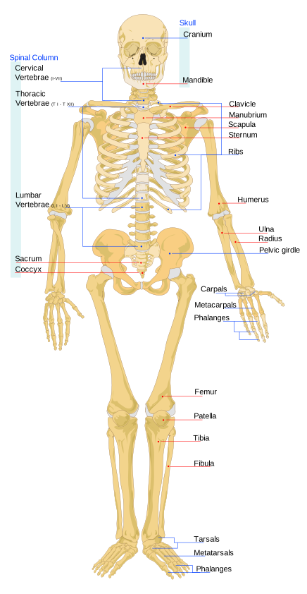
The human skeletal system. Image: LadyofHats (Mariana Ruiz), Public Domain, via Wikimedia Commons.
- 206 bones in adult human body
- Axial skeleton (80 bones): skull, vertebral column (33 vertebrae: 7C+12T+5L+5S+4Coccyx), ribcage
- Appendicular skeleton (126 bones): pectoral girdle, pelvic girdle, limb bones
- Bone composition: organic (collagen) + inorganic (hydroxyapatite = calcium phosphate)
Joints
- Fibrous (sutures in skull): immovable
- Cartilaginous (vertebrae): slightly movable
- Synovial: freely movable; synovial fluid for lubrication
- Types of synovial joints: ball and socket (hip, shoulder), hinge (knee, elbow), pivot (atlas-axis), gliding, saddle
Disorders
- Osteoporosis: reduced bone density; common in post-menopausal women
- Arthritis: joint inflammation; rheumatoid (autoimmune), osteoarthritis (wear and tear)
- Muscular dystrophy: genetic muscle wasting
- Tetany: low Ca2+ causes muscle spasms
Microscopic Structure of Skeletal Muscle
- A muscle is made of bundles of muscle fibres; each fibre contains many parallel myofibrils, and each myofibril is a chain of repeating sarcomeres
- Myosin (thick filament) has a globular head with ATPase activity that can bind actin and ATP; actin (thin filament) has tropomyosin wound around it with troponin complex attached at intervals, which in the resting state blocks the myosin-binding sites
- Light and dark bands: A band (dark, contains thick myosin filaments) and I band (light, contains only thin actin filaments) give skeletal muscle its striated appearance
- Red (slow-twitch) fibres: rich in myoglobin and blood capillaries, depend on aerobic respiration, fatigue slowly (used for sustained activity); white (fast-twitch) fibres: less myoglobin, depend more on anaerobic glycolysis, contract quickly but fatigue fast
Classification of Joints
- Fibrous joints (synarthrosis): immovable; bones held by dense fibrous connective tissue, as in skull sutures
- Cartilaginous joints (amphiarthrosis): slightly movable; bones joined by cartilage, as between adjacent vertebrae (intervertebral discs) and the pubic symphysis
- Synovial joints (diarthrosis): freely movable; bone ends enclosed in a fluid-filled synovial cavity that allows smooth, wide-ranging motion; this is the most common joint type in the limbs
Disorders of the Muscular and Skeletal System
- Myasthenia gravis: autoimmune disorder where antibodies attack acetylcholine receptors at the neuromuscular junction, causing fatigue and weakening of skeletal muscle
- Muscular dystrophy: group of inherited (genetic) diseases causing progressive degeneration of skeletal muscle
- Gout: accumulation of uric acid crystals in joints, causing inflammation and pain
- Rickets: vitamin D deficiency in children leading to soft, deformed bones
Structure of the Contractile Proteins
- Actin (thin filament): two strands of polymerised F-actin, each F-actin being a chain of monomeric G-actin subunits, wound helically together
- Two filaments of the protein tropomyosin run alongside the actin, and a troponin complex sits at regular intervals; in the resting state troponin masks the myosin-binding (active) sites on actin
- Myosin (thick filament): a polymer of many myosin (meromyosin) molecules, each with a globular head plus a short arm projecting outward, and a tail forming the filament backbone
- The projecting head has an ATP-binding site with ATPase activity and an actin-binding site; the head forms the cross-bridge that attaches to actin during contraction
Neuromuscular Junction and Excitation-Contraction Coupling
- A motor neuron and the muscle fibres it supplies form a motor unit; the junction between the neuron's terminal and the muscle fibre membrane (sarcolemma) is the neuromuscular junction / motor end plate
- A nerve impulse causes release of the neurotransmitter acetylcholine, which generates an action potential in the sarcolemma
- The action potential spreads inward along the T-tubules, triggering the sarcoplasmic reticulum to release stored Ca2+ into the sarcoplasm
- Ca2+ binds troponin, shifting tropomyosin to expose the actin active sites — coupling the electrical excitation to the mechanical contraction; when stimulation stops, Ca2+ is pumped back into the sarcoplasmic reticulum and the muscle relaxes
Muscle Energetics and Fatigue
- The immediate energy source for contraction is ATP; muscle also stores creatine phosphate, which rapidly regenerates ATP during short bursts of intense activity
- For sustained activity, ATP is supplied by aerobic respiration (using O2 stored on myoglobin in red fibres); when demand outstrips O2 supply, anaerobic glycolysis takes over
- Muscle fatigue: during heavy anaerobic work, pyruvate is converted to lactic acid, whose accumulation lowers pH and causes the muscle to tire and cramp
- The extra oxygen needed afterwards to clear the accumulated lactic acid is called the oxygen debt, repaid by continued heavy breathing after exercise stops
Axial and Appendicular Skeleton in Detail
- Skull (22 bones): 8 cranial bones enclosing the brain plus 14 facial bones; the skull sits on the first vertebra (atlas), which articulates with the second (axis) to allow head movement; the middle ear also contains 3 tiny ear ossicles on each side, and the U-shaped hyoid bone lies in the neck
- Vertebral column (26 bones in the adult): 7 cervical, 12 thoracic, 5 lumbar, 1 sacrum (5 fused), and 1 coccyx (4 fused); it protects the spinal cord and supports the head
- Rib cage: 12 pairs of ribs attached to the thoracic vertebrae behind; the first 7 pairs are true ribs (joined to the sternum), pairs 8-10 are false ribs (joined indirectly), and pairs 11-12 are floating ribs (not attached to the sternum)
- Appendicular skeleton: the limb bones plus two girdles — the pectoral girdle (each half is a clavicle and a scapula bearing the glenoid cavity for the arm) and the pelvic girdle (each hip bone formed by fused ilium, ischium, and pubis, meeting at the acetabulum that receives the thigh bone)
🚀 NEET Advanced Edge
Why rigor mortis sets in after death: ATP is required to break the actin-myosin cross-bridge (detachment), NOT to form it. After death, ATP production stops, so cross-bridges that have already formed cannot release — leaving muscles permanently contracted/stiff until decomposition breaks down the proteins, the basis of estimating time of death.
Distinguishing band changes precisely: A common trick question gives a diagram and asks which band/zone changes during contraction — only I band and H zone shrink (regions where filaments DON'T overlap, or only thin filament, shrink as overlap increases); the A band (thick filament length) never changes, since myosin filaments themselves don't shorten, only their degree of overlap with actin does.
Myasthenia gravis mechanism in detail: Autoantibodies block/destroy acetylcholine receptors at the neuromuscular junction, so even though the motor neuron releases normal acetylcholine, the muscle fibre cannot respond properly — connecting this disorder directly back to synaptic transmission concepts from the Nervous System chapter.