🎯 Key Points
- Spermatogenesis: spermatogonia→primary spermatocyte→(meiosis I)→secondary spermatocyte→(meiosis II)→spermatid→spermatozoa; Sertoli cells nourish, Leydig cells make testosterone
- Oogenesis: starts BEFORE birth, arrests in meiosis I prophase until puberty; meiosis II completes only AFTER fertilisation (not before)
- Menstrual cycle (28 days): Menstrual(1-5)→Follicular(FSH↑,oestrogen↑)→Ovulation(day14, LH surge)→Luteal(progesterone, corpus luteum)
- hCG maintains the corpus luteum during early pregnancy (basis of pregnancy tests); fertilisation occurs in the fallopian tube; implantation occurs 6-7 days later
- Parturition triggered by oxytocin (posterior pituitary) via the Ferguson reflex — positive feedback loop (contractions→more oxytocin→stronger contractions)
Male Reproductive System
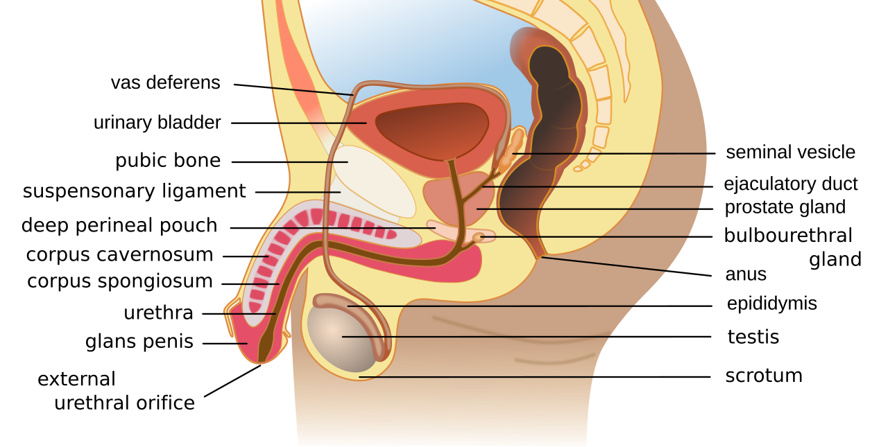
The human male reproductive system: sperm formed in the testis mature in the epididymis and travel via the vas deferens, mixing with secretions of the seminal vesicle, prostate and bulbourethral glands. Image: Wumingbai, CC BY-SA 4.0, via Wikimedia Commons.
- Testes (in scrotum; lower temp needed for sperm): produce sperm and testosterone
- Spermatogenesis: spermatogonia → primary spermatocyte → secondary spermatocyte → spermatid → spermatozoa (meiosis)
- Epididymis: sperm maturation and storage; vas deferens: carries sperm
- Accessory glands: seminal vesicles (fructose), prostate gland (enzymes), Cowper's gland (lubrication)
- Structure of sperm: head (nucleus + acrosome with enzymes), midpiece (mitochondria for energy), tail (flagellum)
Female Reproductive System
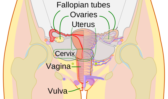
The human female reproductive system. Image: CDC and Mysid, Public Domain, via Wikimedia Commons.
- Ovaries: produce eggs and hormones (oestrogen, progesterone)
- Oogenesis: begins before birth; primary oocyte arrested in meiosis I; resumes at puberty; one egg per month
- Oviduct (fallopian tube): site of fertilization; cilia sweep egg toward uterus
- Uterus: implantation site; endometrium (lining) thickens under progesterone
Menstrual Cycle (28 days)
- Day 1-5: menstruation (endometrium shed)
- Day 6-13: follicular phase (FSH → follicle grows, oestrogen rises)
- Day 14: ovulation (LH surge triggers egg release)
- Day 15-28: luteal phase (corpus luteum secretes progesterone)
- If no fertilization: corpus luteum degenerates, progesterone falls, menstruation begins
Fertilization and Development
- Fertilization: in fallopian tube; acrosome reaction; cortical reaction prevents polyspermy
- Zygote → cleavage → morula → blastocyst → implantation (6-7 days after fertilization)
- Embryonic layers: ectoderm (skin, nervous system), mesoderm (muscles, bones), endoderm (gut lining)
- Placenta: exchange of nutrients and gases; produces hCG (detected in pregnancy tests)
- Gestation: ~266 days (38 weeks); parturition triggered by oxytocin
Reproductive Health
- Contraception: barrier (condom, diaphragm), hormonal (pill), IUD, sterilization
- STIs: HIV, gonorrhoea, syphilis, hepatitis B, chlamydia
- Infertility treatments: IVF (test-tube baby), IUI, GIFT
Spermatogenesis in Detail
- Occurs in the seminiferous tubules of the testis; spermatogonia (diploid, on the inner wall of tubules) divide mitotically to increase in number, then differentiate into primary spermatocytes
- Primary spermatocyte undergoes meiosis I to form two haploid secondary spermatocytes, which undergo meiosis II to form four haploid spermatids
- Spermiogenesis: spermatids transform into mature, motile spermatozoa (sperm); the final release of sperm from Sertoli cells into the lumen of the seminiferous tubule is called spermiation
- Sertoli cells (within seminiferous tubules) provide nutrition to germ cells; Leydig cells (interstitial cells) in the spaces between tubules synthesise and secrete testosterone
- Hormonal control: hypothalamic GnRH stimulates the anterior pituitary to release LH (acts on Leydig cells to produce androgens) and FSH (acts on Sertoli cells, stimulating factors for spermatogenesis)
Oogenesis in Detail
- Begins during embryonic development: oogonia form inside the fetal ovary and enter meiosis I but arrest at the prophase stage, becoming primary oocytes surrounded by granulosa cells to form a primary follicle, in which a girl is born with thousands of these
- At puberty, only a limited number of primary follicles resume development each cycle; the primary follicle develops into a fully grown secondary follicle and then a Graafian follicle, while the primary oocyte completes meiosis I just before ovulation to form a secondary oocyte and a small first polar body
- The secondary oocyte is released at ovulation (LH surge) and begins meiosis II, but this division is completed only if a sperm penetrates the oocyte, producing the ovum and a second polar body
Hormonal Regulation of the Menstrual Cycle
- Menstrual phase: falling progesterone causes the endometrium to break down and shed, released as menstrual flow along with blood
- Follicular phase: rising FSH stimulates follicle growth; the growing follicle secretes oestrogen, which stimulates endometrium repair and proliferation
- Ovulation: a surge of LH (LH surge) triggers rupture of the Graafian follicle, releasing the secondary oocyte around day 14
- Luteal phase: ruptured follicle transforms into the corpus luteum, which secretes large amounts of progesterone, essential for maintaining the endometrium for potential implantation; if fertilisation does not occur, the corpus luteum degenerates, progesterone levels fall sharply, triggering the next menstruation
Hormonal Control of Pregnancy and Parturition
- The developing placenta secretes human chorionic gonadotropin (hCG), which maintains the corpus luteum so it continues producing progesterone needed to sustain pregnancy (hCG is the hormone detected in pregnancy tests)
- The placenta also produces human placental lactogen (hPL), oestrogen, and progesterone in increasing amounts as pregnancy advances, along with relaxin near term
- Parturition (childbirth) is induced by a complex neuroendocrine mechanism: signals originate from the fully developed fetus and the placenta, triggering mild uterine contractions called the "ferguson reflex"; this stimulates release of oxytocin from the maternal posterior pituitary, which acts on uterine muscle to cause strong contractions, leading to expulsion of the baby
Fertilisation and Implantation
- Fertilisation occurs in the ampullary region of the fallopian tube (near the ampullary-isthmic junction); only sperm reaching here can fertilise the ovum
- Acrosomal reaction: enzymes from the sperm acrosome dissolve the zona pellucida and corona radiata; sperm entry then triggers the cortical reaction in the ovum, blocking polyspermy so only one sperm fertilises
- Sperm entry induces the secondary oocyte to complete meiosis II, forming the ovum and a second polar body; the haploid male and female nuclei fuse to form the diploid zygote
- Cleavage: the zygote undergoes mitotic divisions (2, 4, 8, 16 cells) forming a solid morula, which develops into a hollow blastocyst (outer trophoblast + inner cell mass)
- Implantation: the blastocyst attaches to and becomes embedded in the endometrium about 6-7 days after fertilisation
Placenta: Structure and Functions
- The placenta is the structural and functional link between the fetus and the maternal uterine wall, formed by chorionic villi (from the trophoblast) interdigitating with uterine tissue
- Functions: supplies oxygen and nutrients to the fetus and removes carbon dioxide and excretory wastes; the fetus is connected to it by the umbilical cord
- Endocrine role: secretes hormones including hCG, human placental lactogen (hPL), oestrogens and progestogens, and relaxin (later in pregnancy)
- Maternal and fetal blood do NOT normally mix; exchange occurs across the placental membrane by diffusion
Embryonic Development and Pregnancy
- The inner cell mass differentiates into three germ layers — ectoderm (skin, nervous system), mesoderm (muscle, bone, blood, kidney) and endoderm (gut lining, liver, lungs)
- First trimester (months 1-3): most major organs form; the heart forms by about the first month and limbs and external genitalia develop by ~12 weeks
- Second trimester (months 4-6): body hair and eyelashes appear; fetal movements and heartbeat can be detected
- Third trimester (months 7-9): rapid growth and organ maturation; full gestation is about 9 months (roughly 266-280 days)
Parturition and Lactation
- Parturition (childbirth): vigorous uterine contractions driven by the oxytocin-mediated Ferguson reflex (positive feedback) expel the fetus; the fully developed fetus and placenta generate the initial triggering signals
- Lactation: the mammary glands enlarge and begin milk production near the end of pregnancy under prolactin, while oxytocin causes milk ejection (the let-down reflex)
- Colostrum: the yellowish first milk secreted in the initial days after birth is rich in antibodies (IgA) and gives the newborn passive immunity — feeding it is strongly recommended
Male Duct System and Semen
- Sperm made in the seminiferous tubules travel through the rete testis → vasa efferentia → epididymis (where they mature and are stored) → vas deferens → ejaculatory duct → urethra, which opens at the tip of the penis
- Each testis lies in the scrotum, which keeps testicular temperature about 2-2.5°C below body temperature — essential for normal spermatogenesis
- Internally each testis is divided into around 250 compartments (testicular lobules), each containing the highly coiled seminiferous tubules lined by spermatogonia and Sertoli cells
- Semen = sperm suspended in the secretions of the accessory glands; seminal vesicle fluid (rich in fructose that fuels sperm) forms the bulk, while the prostate and Cowper's (bulbourethral) glands add enzymes and lubricating fluid
Female Reproductive Ducts and Accessory Organs
- Each fallopian tube (oviduct) has, running from the ovary inward: the funnel-shaped infundibulum bearing finger-like fimbriae that collect the ovulated ovum, the wider ampulla (the usual site of fertilisation), and the narrow isthmus that joins the uterus
- The uterus (womb) wall has three layers: an outer perimetrium, a thick muscular middle myometrium (source of the strong contractions of childbirth), and an inner glandular endometrium that undergoes cyclic changes through the menstrual cycle
- The uterus opens into the vagina through a narrow cervix; the cervical canal and vagina together form the birth canal
- The mammary glands are paired accessory glands whose glandular tissue is organised into mammary lobes containing clusters of alveoli; milk secreted into the alveoli passes via mammary tubules → mammary ducts → mammary ampulla → lactiferous duct to the nipple
Menarche and Menopause
- Reproductive (menstrual) cycles begin at puberty; the very first menstruation is called menarche
- Cycles then recur roughly every 28-29 days throughout the reproductive years, pausing during pregnancy
- Menopause is the permanent cessation of menstrual cycles, usually around 45-50 years of age, after which the woman can no longer reproduce
- Cyclic menstruation is a distinctive feature of primates (apes, monkeys and humans) and is an indicator of normal reproductive health; its absence may signal pregnancy, stress, or an underlying condition
🚀 NEET Advanced Edge
Why oogenesis pauses TWICE but spermatogenesis doesn't: Oogenesis arrests once before birth (meiosis I, until puberty) and again after ovulation (meiosis II, until fertilisation) — these checkpoints conserve the egg's resources and ensure meiosis II only completes if fertilisation actually happens, preventing wasted completion of an unfertilised egg. Spermatogenesis runs continuously from puberty onward with no such arrest.
The Ferguson reflex as positive feedback: Unlike most physiological control loops (negative feedback, self-correcting), parturition is a rare POSITIVE feedback loop: uterine stretch → oxytocin release → stronger contractions → more stretch → even more oxytocin — escalating until delivery, then the loop is broken by the baby's birth itself.
Worked reasoning: A pregnancy test detects hCG in urine. Why does hCG appear so early and specifically signal pregnancy? Because hCG is produced ONLY by the developing placenta/trophoblast (not by the ovary or any other tissue), and it rises quickly after implantation to rescue the corpus luteum from degenerating — making it both an early AND a highly specific marker.